Skip to content


A rhinoplasty simulation is best used as a starting point for a conversation, not a promise of a finished result. Computer imaging for rhinoplasty digitally adjusts the nasal contour in a photograph, helping you and your surgeon discuss possible changes and how they may relate to your facial features. It can illustrate an aesthetic direction, but it can’t guarantee the surgical outcome.
It’s natural to want a clearer picture before making a decision. This article explains what a simulation can show, how it can support communication with your surgeon, and why it should be considered alongside, not instead of, a discussion of nasal function. It also covers how your preferences can help shape a personalized plan and what an image can’t predict about surgery and healing.
Key Takeaways
- Use a simulation to express the nasal changes you’re considering and discuss how they relate to your facial features.
- Understand what a still image can show, and why it can’t predict every angle or the effects of surgery and healing.
- Bring appearance goals and nasal function into the same conversation when discussing a personalized surgical plan.
- Learn how a surgeon can use photographs and imaging during consultation to make aesthetic goals more concrete.
What Is Computer Imaging in a Rhinoplasty Consultation?
Computer imaging for rhinoplasty digitally adjusts the nose in a patient photograph. In the United States, the cost depends on the case and is given after a quote request; factors include the planning involved and whether imaging is part of the consultation.
Consultation Communication and Surgical Planning
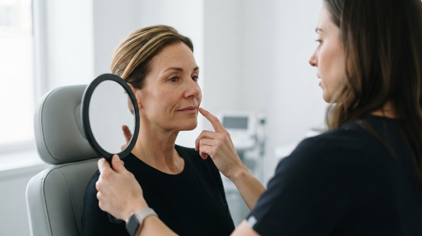
A simulation gives the surgeon and patient a visual reference for discussing possible nasal changes. For example, an edited profile can help clarify whether you prefer a smoother bridge or a different tip contour. This shared image makes aesthetic preferences more specific than verbal descriptions alone.
Your surgeon can use that discussion to develop a surgical plan around your goals and facial features. The image can also highlight when a requested change needs further discussion. It is a planning reference, not a surgical instruction or a promise of a particular result.
Personalizing the Surgical Plan
An individualized plan considers your facial features and the changes you want to discuss. Imaging can illustrate adjustments to nasal length, width, or nostril contour, separately or together. For example, if you’re concerned about both width and tip shape, you can compare how those goals appear in one edited photograph.
Your surgeon can use this visual comparison to focus the discussion on priorities. You might prefer a subtle bridge change over a narrower overall appearance. Clarifying that distinction connects your personal preference with a plan tailored to you, rather than treating one image as the right nose for everyone.
Limits of Predicted Results
A simulation illustrates a proposed appearance, but it can’t guarantee an exact surgical result. A photograph captures one angle, and a two-dimensional edit may not show the nose from other viewpoints. For instance, a profile image alone cannot fully convey how a proposed tip change looks from the front.
Surgical technique, tissue response, and healing also affect the final appearance. Even a carefully created image may depict a change that is difficult or impossible to reproduce surgically. Discussing these limits keeps the simulation in perspective: it supports planning and communication, while your surgeon’s assessment guides what is feasible.
How can a rhinoplasty simulation support communication?
A simulation gives the surgeon and patient a visual reference for discussing possible changes. In the United States, rules may differ between private consultations and public advertising. How an image is presented and what claims accompany it matter.
Photographs a Surgeon May Use for Rhinoplasty Simulation
Photographs provide the starting point for a simulation. A surgeon may digitally edit a patient photo or trace proposed changes over it. The photo shows only its captured angle, so a profile view cannot show the full frontal appearance. Some workflows ask patients to upload five photographs from set angles after booking, so a simulation can be prepared. Comparing views helps clarify what each image can show.
Discussing Simulations During Consultation
During consultation, a simulation can help you describe your preferences more clearly. You might indicate a preferred tip shape on an edited image, then discuss how that change relates to other views. The image can focus the conversation, but it cannot show how surgery or healing will affect your nose.
The Authors of the randomized rhinoplasty study reported on 44 Asian patients having rhinoplasty with rib cartilage; imaging improved satisfaction with doctor-patient communication but didn’t change satisfaction with the outcome. This finding highlights the image’s role as a discussion aid, not a promise of results.
Planning Surgical Modifications
Your surgeon can use a simulation to discuss proposed modifications before developing a surgical plan. For example, you can compare a subtle bridge adjustment with a more noticeable change. You can also discuss which features matter most, rather than treating every visible edit as a surgical instruction. This connects a visual preference with a considered plan.
Computer imaging for rhinoplasty can show an intended contour, but it can’t fully model surgical technique, tissue response, or healing. A digital edit may also depict a change that is technically unrealistic or surgically impossible. Discussing these limits alongside each proposed modification helps distinguish an aesthetic reference from a predictable postoperative photograph.

How do aesthetic goals and nasal function fit together?
A rhinoplasty plan considers the appearance you want alongside nasal function. Computer imaging can help make your aesthetic preferences more concrete for discussion.
Communicating Cosmetic Goals
A simulation turns a general preference into a visible point of discussion. For example, “a softer-looking nose” can mean different changes to different people. An edited image helps clarify whether you mean a change to nasal length, width, or nostril contour. You can respond to the proposed shape and explain which details matter most. This shared reference helps your surgeon understand your aesthetic priorities.
These preferences guide the discussion, not a fixed template. A particular image can prompt questions about how one change affects the overall appearance. Your surgeon can use your feedback to refine the conversation and consider your facial features alongside the requested change.
Balancing Appearance and Nasal Function
Aesthetic goals and nasal function belong in the same planning conversation. If appearance is your main concern, describe the specific change you hope to see. If breathing is also a concern, explain that clearly during your consultation. These priorities give your surgeon a fuller understanding of what you want addressed.
Imaging can illustrate proposed changes to the external nasal shape, but it doesn’t establish how your nose functions. For example, a narrower-looking contour in a simulation doesn’t show whether breathing will improve or remain comfortable. Your surgeon considers your preferences, facial aesthetics, and functional concerns together when developing a personalized plan.
Assessing Realistic Goals
A simulation can help you compare a desired change with the features shown in your photograph. For instance, you and your surgeon might discuss whether a proposed alteration looks balanced with your other facial features. This visual comparison gives you a specific way to describe what you like, what feels excessive, and which details you’d reconsider.
Use the image to clarify priorities, not to set an exact outcome. A digital contour cannot fully represent surgical technique, tissue response, or healing. Your surgeon’s discussion of feasibility helps distinguish an aesthetic preference from a practical surgical goal. That distinction can shape a plan that respects both appearance and function.
How computer imaging works during consultation
During consultation, imaging turns a photograph into a visual planning aid. Surgeons may use edited photographs, traced images, anatomical diagrams, or three-dimensional visualization. No single tool has been shown to be better.
The Consultation Imaging Process
The process begins with photographs that show the nose from selected angles. Your surgeon can then adjust the nasal contour digitally or trace a proposed change over a photograph. For example, a profile view can illustrate a bridge adjustment. The image gives you both a concrete reference for discussing the intended change.
Next, you and your surgeon review the proposed appearance together. You can describe what aligns with your goals and what does not. With a two-dimensional image, discussion is limited to the angle shown. Some three-dimensional methods add views of facial contours from multiple viewpoints, offering a broader visual reference.
Software and Imaging Technology
Rhinoplasty visualization can involve computer software, traced photographs, or other visual aids. The approach varies, and no particular software is needed to make an image useful. What matters is whether the method helps clarify the proposed change and supports a focused discussion about your goals. In clinical settings where visual precision is essential, Synergy Medical Inc. provides guidance on evaluating medical-grade imaging displays to support detailed anatomical review.
A two-dimensional simulation modifies a photograph but cannot display every angle. Three-dimensional visualization can provide additional perspectives, though it still cannot model every surgical or healing factor. A photograph captures one view, not a surgical change. Treat each image as a discussion tool, not a complete representation of your nose after surgery.
Use the consultation to clarify what the image represents and how it informs the plan. Discuss rhinoplasty planning in relation to your aesthetic goals and nasal function.
Dr. Leonard Grossman: Rhinoplasty
Dr. Leonard Grossman offers rhinoplasty for patients considering changes to nasal appearance. If a simulation has helped clarify your preferences, the next step is discussing those goals alongside facial features and nasal function. A visual preview can guide that conversation, while the surgical plan considers what is appropriate for your individual anatomy and priorities.
Bring the changes you’re considering into the discussion, such as a different nasal bridge contour or tip shape. Explain which features matter most and whether breathing is also a concern. This gives your consultation a clear starting point without treating a digital image as a guaranteed result.
If you’re ready to discuss your goals and questions, contact Dr. Leonard Grossman.
Take the Next Step Toward a Clearer Plan
Use the ideas from your simulation to guide a thoughtful consultation. Before meeting with a surgeon, consider which changes matter most to you and whether nasal function is also part of your goals. This reflection can help you explain your priorities and make the conversation more specific.
Computer imaging for rhinoplasty can help you express an aesthetic direction, but the most useful next step is discussing how it fits your individual needs. Dr. Leonard Grossman also offers Fat Grafting to the Face, Fat Grafting to the Breasts, and Fat Grafting to the Buttocks as separate aesthetic procedures.
If you’re ready to talk through your rhinoplasty goals, contact Dr. Leonard Grossman to discuss rhinoplasty. A considered conversation can help you move forward with greater clarity and confidence.
Frequently Asked Questions
Can I See What I Would Look Like After Rhinoplasty?
Yes, a surgeon may show you a digitally adjusted photograph to illustrate a possible nasal change. In a consultation, computer imaging for rhinoplasty can help you compare visual options, such as a more subtle or more defined contour. The image is a planning aid, not a photograph of your actual future appearance. Use it to explain which features feel closer to your goals.
Can AI Rhinoplasty Simulations Used in a Consultation Show Me How I Might Look?
An AI-generated simulation may create a visual mock-up of a possible nasal change, but it can’t establish your surgical result. Ask what the image represents and which parts were digitally altered. For example, if software changes the nose while leaving the rest of the photograph untouched, the image may help you discuss appearance, but it won’t show how surgery affects surrounding tissues or healing.
Does a rhinoplasty simulation guarantee the result I want?
No. A simulation can’t guarantee that surgery will produce the exact shape shown. It represents an aesthetic idea, while the surgical plan must account for your anatomy and what can be achieved through surgery. If an edited image suggests a dramatic change, discuss which parts are realistic and which may need adjustment. That conversation helps set clearer expectations before you decide on surgery.
How Close Does the Simulation Get to the Real Result?
There’s no single accuracy rate that predicts how closely an individual result will match a simulation. In one study involving 38 rhinoplasty subjects, expert judges rated the imaging system’s predictive accuracy 2.98 on a 5-point scale. They compared postoperative photographs with computer predictions. That rating offers context, not a personal forecast, so use your own simulation as a discussion reference.
What additional perspective can three-dimensional imaging provide?
Three-dimensional visualization can show facial contours from multiple viewpoints, rather than limiting discussion to one edited photograph. This may help you consider how a proposed change appears beyond a single profile or front view. It still doesn’t establish how your nose will look after surgery. Use the additional perspectives to identify questions about shape and facial balance.
What can imaging help you understand before surgery?
Imaging can help you clarify which visual changes matter most before making a decision. For example, comparing two proposed shapes may reveal that you prefer one feature but reject another. It can also help you prepare focused questions about how a proposed change relates to your goals. The image won’t answer every surgical question, but it can make your preferences easier to discuss.